Acne Scars vs Dark Spots vs PIH — What You Actually Have and How to Fade Each One (2025)
The flat brown mark a pimple leaves behind is not a scar. The sun-triggered blotch on your cheek is not PIH. And the textured indent that catches light at an angle is not something a serum can fill. These three conditions have completely different biology, completely different timelines, and require completely different treatments. Using the wrong one wastes months. This guide tells you exactly which one you have — and what actually works for it.

The Indian skin care market is flooded with products that promise to "fade acne marks" — but that phrase covers three entirely distinct biological conditions that have nothing in common except that they follow a breakout or skin injury. The market's failure to distinguish them is, in large part, why so many people cycle through product after product with minimal results: they are treating the wrong thing.
Post-inflammatory hyperpigmentation (PIH) is a pigmentation problem — an excess of melanin deposited in the skin after inflammation. The skin surface is smooth. The damage is in the pigment system. This responds to topical depigmenting agents: Vitamin C, Niacinamide, Alpha Arbutin, Tranexamic Acid, AHAs. But only if sun protection is consistent — without SPF, every topical is fighting a losing battle against continuous UV-driven melanocyte re-stimulation.
Dark spots from other causes (solar damage, melasma, friction marks) share PIH's flat surface but have different triggers, different depths, and different treatment responses. Melasma, in particular, is frequently misidentified as PIH and treated with the same products — with far less success, because melasma is hormonally driven and requires a different protocol.
Acne scars are a structural problem — physical damage to the collagen architecture of the dermis. The surface is not smooth. Topical depigmenting agents do nothing for depressed collagen voids. This requires collagen remodelling: retinoids, chemical peels, microneedling, or professional procedures depending on depth and type.
The first and most important step: correctly diagnosing which one — or which combination — you are dealing with.
⚡ PIH vs Dark Spots vs Acne Scars — Quick Diagnostic Reference
| Feature | PIH | Dark Spots (Sun / Melasma) | Acne Scars |
|---|---|---|---|
| Texture | Completely flat — smooth to touch | Flat — smooth to touch | Has texture — depressed, raised, or pitted |
| Cause | Inflammation (acne, wound, eczema, waxing, friction) | UV exposure (sun spots), hormones (melasma), friction or ageing | Deep dermis damage during active acne (collagen destruction or overproduction) |
| Colour | Pink/red (fresh) → brown/dark brown → fades over months | Light to dark brown; melasma often has irregular border; solar spots have sharp edges | Skin-coloured, pink, or brown (colour independent of depth) |
| Location pattern | Exactly where the pimple was; follows breakout map | Sun-exposed areas (cheeks, forehead, upper lip for melasma); areas of chronic friction | Where acne was most severe — cheeks, jaw, forehead |
| Worsens in sun? | Yes — significantly | Yes — especially melasma | Scars themselves do not darken; any associated PIH does |
| What works | Vitamin C, Niacinamide, Alpha Arbutin, AHAs, Tranexamic Acid + SPF | Same as PIH for superficial; Hydroquinone / Azelaic Acid for melasma; SPF essential | Retinoids, microneedling, chemical peels, fillers, laser — depending on type |
| What doesn't work | Scar fillers, microneedling (not for flat PIH) | Spot treatments not matched to trigger or depth | Depigmenting serums (cannot fill collagen voids) |
| Timeline to fade | 3–12 months with treatment + SPF | Superficial: 3–12 months. Deep melasma: long-term management | Rolling: 6–18 months. Boxcar/ice pick: professional treatment required |
The 4-Step Diagnosis — Which One Do You Actually Have?
Most people have a combination — active PIH from recent breakouts overlying older textured scars, or melasma in the same area as PIH from inflammation. The diagnostic process separates these layers so you can address each correctly.
Run a clean fingertip gently across the area of concern in good side-lighting or at a slight angle to the light. This is the single most definitive test between scars and pigmentation.
- Completely smooth, no variation in surface level: You have pigmentation — PIH, dark spot, or melasma. No textural component means no structural scar.
- Slight depression or indent: Atrophic scar — collagen was destroyed during the inflammation. The type (ice pick, boxcar, rolling) determines severity.
- Raised, firm area: Hypertrophic scar or keloid — excess collagen was produced during healing. Common on the jaw, chest, and shoulders in people with a genetic tendency toward raised scars.
- Mix of smooth discolouration AND textured areas: You have both PIH and scars — common in moderate to severe acne histories. Address each with its own targeted treatment.
Trace the origin of each mark. The trigger determines the type:
- Appeared exactly where a pimple or wound was: PIH. The inflammation from the breakout triggered excess melanin production in that precise area.
- Appeared on sun-exposed areas without a preceding pimple: Solar lentigo (sun spot) — UV-driven melanocyte activation independent of inflammation. Common on cheeks, nose, forehead, upper lip in Indian adults over 25.
- Appeared symmetrically across cheeks, forehead, or upper lip, worsened in summer, worsened during or after pregnancy or hormonal contraceptive use: Melasma — hormonally driven overactivation of melanocytes. Melasma is not PIH and does not respond to the same degree to the same treatments.
- Appeared in areas of chronic friction (between thighs, underarms, neck folds): Friction-driven hyperpigmentation — mechanically triggered melanocyte activation. Requires reducing friction first; topicals alone are insufficient if the friction continues.
- Permanent change in surface texture at the exact site of a past severe pimple: Acne scar — dermis damage during a cyst or nodule that the inflammation was too deep and severe to heal without structural loss.
The colour of flat marks gives you clues about depth — which determines treatment response:
- Pink or red flat marks: Recent PIH — the inflammation has recently resolved and the excess melanin is still in the epidermis (outermost layer). This responds fastest to topical treatment — typically within 3–6 months.
- Light brown, tan: Epidermal pigmentation (PIH or solar spot) — responds well to topical brightening actives and AHAs that accelerate epidermal turnover.
- Dark brown to near-black: May be deep epidermal or dermal pigmentation — treatment response is slower. Deeper melasma in this colour range responds poorly to topicals alone.
- Greyish or bluish tint in brown skin: Dermal pigmentation — melanin is deposited in the dermis, not just the epidermis. Topicals reach the epidermis effectively but have limited penetration to the dermis. This typically requires professional treatment (chemical peels, laser) for meaningful improvement.
The Wood's Lamp test: A UV Wood's lamp held close to the face in a darkened room enhances epidermal pigmentation (it appears more pronounced under UV) but does not enhance dermal pigmentation (it looks the same or disappears). This is the clinical standard for assessing pigmentation depth. Dermatologists routinely use this to determine whether a patient's hyperpigmentation will respond to topicals. If you have access to a consultation, this single test saves months of ineffective product trials.
Observe whether your marks change with seasons or sun exposure — this confirms the type and guides urgency of sun protection.
- Marks consistently darker in summer / after sun exposure: Active UV-driven pigmentation — melanocytes in the affected area are UV-sensitive. PIH, solar spots, and melasma all behave this way. SPF 50+ is not optional — it is the primary treatment vehicle, without which all other interventions are partially reversed by daily sun exposure.
- Marks stable across seasons, unchanged by sun: Suggests structural scarring rather than active pigmentation. The surface texture change is physical (collagen void), not driven by melanocyte activity.
- Marks dramatically worse during summer even without new breakouts: Strong indicator of melasma rather than PIH — melasma's UV sensitivity is extreme and its summer darkening is typically much more dramatic than PIH from old acne.
Post-Inflammatory Hyperpigmentation (PIH) — Biology, Causes, and Treatment

What PIH Actually Is
PIH is an excess melanin response to skin inflammation. When skin is injured — by acne, a cut, eczema, waxing trauma, or even aggressive scrubbing — the keratinocytes (skin cells) release inflammatory signals including prostaglandins and cytokines. These signals stimulate nearby melanocytes to produce more melanin as a protective response. This melanin is then transferred to surrounding keratinocytes and deposited in the epidermis — or, in severe inflammation, in the dermis — leaving a visible discolouration long after the primary inflammation has resolved.
PIH is significantly more pronounced in darker skin tones (Fitzpatrick IV–VI, which encompasses most Indians) because higher baseline melanin levels mean a stronger and longer-lasting pigmentation response to the same inflammatory stimulus. A pimple that leaves a barely-visible mark on light skin can leave a dark, persistent mark on Indian skin — this is not abnormal, it is a predictable biological difference, not a skin "problem."
The key clinical fact: PIH is not permanent by nature. The pigment in epidermal PIH turns over with normal skin cell cycling — the epidermis renews completely every 28–40 days. Without UV reinforcement and without new inflammation, PIH would gradually fade on its own. The problem is that UV exposure continuously restimulates melanocytes in the affected area, and new breakouts add new PIH on top of old marks — making the condition appear chronic when it is actually continuously being renewed.
What Works for PIH — The Evidence-Ranked Treatment Hierarchy
Tier 1 — Non-Negotiable Foundation: SPF 50+ Sunscreen Daily
No topical brightening agent works at full efficacy without consistent sun protection. UV exposure re-stimulates the melanocytes in PIH-affected areas daily — every unprotected hour in the Indian sun adds new pigment that any evening serum must then work harder to clear. Broad-spectrum SPF 50+ with PA++++ (UVA protection critical for pigmentation, not just burn prevention) applied every morning, reapplied every 2–3 hours outdoors. This is not optional; it is the single intervention with the highest impact on PIH fading speed. Using a Vitamin C serum without sunscreen is significantly less effective than sunscreen alone.
Indian context: India's UV Index regularly exceeds 10 from March to October in most cities — equivalent to extreme UV. Indoor exposure through windows also transmits UVA. SPF is a year-round morning step regardless of season or whether you plan to be outdoors.
Tier 2 — Active Depigmentation: Vitamin C + Niacinamide
Vitamin C (L-Ascorbic Acid, 10–20%): Inhibits tyrosinase — the key enzyme in the melanin synthesis pathway — reducing new melanin production. Also provides antioxidant protection against UV-driven oxidative stress that triggers melanocyte activity. Most effective in the 10–20% concentration range at pH below 3.5; above pH 3.5 it degrades rapidly. Applied in the morning before SPF for synergistic UV protection.
Niacinamide (5–10%): Inhibits melanosome transfer — the process by which melanin pigment packets move from melanocytes to surrounding skin cells. It does not stop melanin production; it stops it from being distributed. Well-tolerated by Indian skin, no sensitisation, can be used morning and evening. Combined with Vitamin C, addresses two separate steps in the pigmentation pathway — a complementary, not redundant, pairing. See the Niacinamide guide and Vitamin C guide for full evidence reviews.
Tier 3 — Accelerate Cell Turnover: AHAs and Retinoids
AHAs (Glycolic Acid, Lactic Acid): Accelerate epidermal shedding, speeding up the natural process by which pigmented skin cells are shed and replaced by fresh, un-pigmented cells. Glycolic acid (smallest molecular size, deepest penetration) at 5–10% used 2–3× per week; Lactic Acid (larger molecule, gentler, more hydrating) at 5–10% for sensitive skin. AHAs exfoliate the PIH away faster than natural cell turnover allows — dramatically reducing the 6–12 month timeline to 3–4 months in consistent use.
Retinoids (Retinol 0.1–0.5%, Adapalene 0.1%): Accelerate skin cell turnover at a deeper level than AHAs, normalise melanin distribution, and — critically — treat the active acne that is generating new PIH. Adapalene 0.1% is available over the counter in India (Differin) and is the best-evidenced retinoid for simultaneous acne treatment and PIH reduction. Start slowly (1–2× per week), use only at night, always follow with SPF the next morning.
Do not combine AHAs and Retinoids in the same routine step — the combined pH disruption and irritation increases the risk of new inflammation, which creates new PIH. Alternate: AHAs on evenings 1 and 3; retinoid on evenings 2 and 4.
Tier 4 — Targeted Actives: Alpha Arbutin and Tranexamic Acid
Alpha Arbutin (1–2%): A glycosylated form of hydroquinone that inhibits tyrosinase without the side effects associated with hydroquinone itself (ochronosis with long-term use, irritation). Effective for epidermal PIH, well-tolerated in Indian skin. Can be used morning and evening. Most effective when layered with Niacinamide for dual mechanism inhibition.
Tranexamic Acid (2–5%): Inhibits keratinocyte-derived plasminogen activator, a signalling molecule that activates melanocytes after UV exposure and inflammation. Particularly effective for melasma and UV-exacerbated PIH. Available in topical form as a serum or in prescription oral form (used short-term by dermatologists for resistant melasma). The topical concentration (2–5%) is safe for long-term use without the systemic effects of the oral form.
Liquorice extract (Glabridin): Traditional Ayurvedic brightening ingredient with documented tyrosinase-inhibition efficacy. See the Liquorice skin guide for the full evidence breakdown.
Dark Spots — Solar Damage, Melasma, and Other Causes
Not every flat discolouration on the face is PIH. Dark spots from UV exposure, hormonal triggers, and chronic friction have different mechanisms and different treatment responses — including significantly lower response rates to the same topicals that fade PIH effectively.
What they are: Well-defined, flat, brown spots that appear on chronically sun-exposed areas — cheeks, nose bridge, forehead, upper lip, décolletage, hands. They result from long-term cumulative UV exposure causing localised proliferation and hyperactivation of melanocytes — permanently altered cells that remain hyperpigmented independently of inflammation. Unlike PIH, they do not follow a past pimple and are not caused by injury.
How to distinguish from PIH: Solar lentigines have sharp, well-defined borders; PIH is usually more diffuse. Solar spots do not correspond to past breakout locations. Solar spots appear predominantly on the upper face (most UV-exposed zones); PIH appears exactly where acne was located.
Treatment: Respond to the same topicals as PIH (Vitamin C, AHAs, Alpha Arbutin) but more slowly — because the melanocyte proliferation is structural rather than a transient inflammation-driven response. Professional treatments (IPL, laser, deep chemical peels) are more efficient for established solar spots than topicals alone. Prevention: SPF 50+ PA++++ applied daily from your twenties is the only intervention that prevents new solar lentigines from forming.
What it is: A chronic, hormonally mediated hyperpigmentation condition characterised by symmetrical brown or greyish-brown patches on the face — predominantly the cheeks, forehead, upper lip, and chin. Melasma is driven by oestrogen and progesterone stimulating melanocytes, making it significantly more common in women during pregnancy ("mask of pregnancy"), while on combined oral contraceptives, or with hormonal IUDs. It is also exacerbated dramatically by UV exposure — UV acts as a co-trigger, amplifying the hormonal melanocyte stimulation.
Why it is harder to treat than PIH: Melasma involves both epidermal and often dermal pigmentation — and the dermal component does not respond to topicals that only reach the epidermis. Melasma also recurs — once the melanocytes are sensitised, they remain reactive to UV and hormonal triggers. Even after successful treatment, re-exposure to the original trigger (sun, hormonal contraceptives, pregnancy) restores the melasma. It is managed, not cured.
Treatment approach: Strict and year-round SPF use (the single most important intervention — without it, no treatment works). Topicals: Tranexamic Acid (most effective for melasma specifically), Niacinamide, Azelaic Acid (15–20%, available by prescription, strong evidence for melasma). Hydroquinone (2–4%, prescription in India) remains the gold standard but requires breaks due to risk of ochronosis with extended use. Professional: chemical peels (glycolic, kojic acid combinations), low-fluence Q-switched laser for dermal component. Discontinuing hormonal contraceptives, where medically appropriate, dramatically improves melasma response to treatment.
What it is: Chronic friction or pressure on skin surfaces activates keratinocytes to release inflammatory mediators — the same pathway as PIH, but triggered mechanically rather than by infection or injury. Common sites in India: inner thighs (from clothing friction), underarms (from hair removal trauma and deodorant), neck folds (collar friction), elbows and knees (pressure on hard surfaces). Also common from tight underwear waistbands and bra straps.
Why it doesn't respond to topicals alone: If the friction source is ongoing — daily clothing contact, regular hair removal, habitual pressure — topical brightening agents are working against a continuously active trigger. The skin is simultaneously being depigmented by the actives and re-pigmented by the friction. Reducing the friction source must happen first or concurrently.
Treatment: Identify and reduce friction: looser clothing, moisture-wicking fabrics in high-friction zones, switching hair removal method (electrolysis or laser over repeated waxing or shaving for underarms and bikini line), applying barrier cream in rub zones before exercise. Then add the same depigmenting actives used for PIH — Niacinamide, AHAs. Results come only after the mechanical trigger is addressed.
Acne Scars — Types, Biology, and What Actually Works
True acne scars are structural — physical damage to the dermis that creates either a collagen deficit (atrophic, depressed scars) or collagen excess (hypertrophic, raised scars). The dermis cannot spontaneously rebuild lost collagen to the same density as undamaged tissue, which is why scars are permanent without intervention. Understanding the specific type of scar is essential because different types require different professional approaches.
Ice Pick Scars — Narrow, Deep, Most Resistant
What they look like: Narrow, V-shaped channels punched into the skin — like a pin was pressed into the cheek. Usually 1–2mm wide, but can extend 3–4mm deep into the dermis. The narrow width makes them appear as tiny pores or skin punctures in diffuse lighting, but in direct side-lighting or flash photography they are clearly visible.
Why they form: Deep, severely inflamed cysts and nodules that extend into the lower dermis destroy collagen at depth. The narrow channel results from the skin attempting to heal from the inside out but lacking enough collagen to fill the void.
What works: TCA CROSS (Chemical Reconstruction of Skin Scars) — highly concentrated TCA (80–100%) applied with a toothpick precisely into the base of each ice pick scar, stimulating collagen production in the channel. Requires multiple sessions spaced 6–8 weeks apart; performed by a dermatologist. Punch excision (surgical removal of the scar channel, suturing closed) for very deep examples. Topical products have no meaningful effect on ice pick scars.
Boxcar Scars — Defined Edges, Moderate to Severe
What they look like: Round or oval depressions with sharp, vertical edges — like a box stamped into the skin. Wider than ice pick scars (1.5–4mm) but with clearly defined borders. Shallow boxcar scars (0.1–0.5mm deep) respond better to treatment; deep boxcar scars (0.5mm+) require more aggressive intervention.
Why they form: Wide column of dermis destroyed by inflammation, leaving a broader void than an ice pick. The sharp walls are formed by fibrous tissue that anchored the surrounding skin as it healed.
What works: Microneedling (professional radiofrequency microneedling or dermaroller) — induces controlled micro-injury that triggers collagen synthesis throughout the scar base, gradually filling the depression over multiple sessions. Medium-depth chemical peels (TCA 20–35%) improve shallow boxcar scars by resurfacing and stimulating collagen. Subcision (inserting a needle under the scar to break fibrous attachments, allowing the surface to rise) combined with fillers for deeper examples. For shallow boxcar scars: at-home dermarolling (0.5–1mm) with consistent retinoid use can produce visible improvement over 6–12 months.
Rolling Scars — Broad, Wave-Like, Most Responsive to Treatment
What they look like: Broad, shallow depressions with sloping, undulating edges — the skin surface has a wave-like or rolling appearance. The skin surface lacks sharp edges; it simply dips gradually and rises again. Most visible in side-lighting. Create a shadow effect in photographs that is disproportionate to how they feel to the touch.
Why they form: Fibrous bands of scar tissue connect the base of the dermis to the underside of the skin surface, tethering it and pulling it downward. Unlike ice pick and boxcar scars which are vertical collagen voids, rolling scars are caused by tethering — not just loss.
What works: Subcision is particularly effective for rolling scars — releasing the fibrous tethers allows the skin surface to rise. Followed by microneedling to build collagen in the now-released area. Rolling scars also respond best to topical collagen stimulators (Retinoids, Vitamin C) of all scar types — the gradual collagen rebuilding addresses the shallow, broad depression. With consistent at-home treatment (retinoid + microneedling 0.5–0.75mm roller, monthly) and professional subcision, significant visible improvement is achievable in 6–18 months.
Hypertrophic Scars and Keloids — Raised, Excess Collagen
What they look like: Raised, firm, often pink or darker-than-skin-tone tissue at the site of healed acne. Hypertrophic scars remain within the original wound boundary; keloids extend beyond it and continue growing over time.
Who gets them: Raised scars are more common in darker skin types (Fitzpatrick IV–VI) — including most Indian skin — and are genetically determined. People who form keloids on the chest, shoulders, or earlobes are at higher risk for them on the face as well.
What works: Silicone gel sheets or silicone scar gels applied consistently over 3–6 months — occlude the scar and regulate hydration, reducing fibroblast activity and excess collagen. Onion extract (allium cepa) gel — reduces scar redness and hardness over time. Intralesional corticosteroid injections (administered by a dermatologist) flatten hypertrophic scars in 2–6 sessions by suppressing excess collagen production. True keloids may require laser (PDL, Nd:YAG) or surgical excision with steroid injection to prevent recurrence.
The Rule That Applies to All Three — Sunscreen Is Not Optional
The Single Biggest Mistake in Indian Skin Care for Discolouration
Using brightening serums without daily SPF 50+ PA++++ is the most common reason Indian skin care routines for hyperpigmentation fail. UV radiation does three things that collectively undo every topical treatment: it directly stimulates melanocyte activity in all hyperpigmented areas (re-darkening marks that were fading), it triggers new inflammation (creating new PIH), and it generates reactive oxygen species (ROS) that oxidise Vitamin C serums and partially deactivate their anti-melanogenic effect before they can work.
SPF use is not a complementary step to brightening actives. It is the primary treatment. In India's UV environment — UV index 10–12 from March to October in most cities, with significant UVA transmitted even indoors through windows — unprotected daily skin exposure continuously reverses the work of a Vitamin C or Niacinamide routine. A patient who uses SPF 50+ PA++++ consistently and nothing else will typically see greater PIH improvement over 6 months than a patient using every brightening active without SPF.
Practical SPF minimum: SPF 50+, PA++++ (4 plus signs = highest UVA protection), broad-spectrum. Applied as the last morning step, after moisturiser. Reapplied every 2–3 hours if outdoors. No exceptions during treatment for hyperpigmentation of any type. See the complete sunscreen guide for Indian skin for how to choose a formula that does not cause white cast on deeper skin tones.
💡 Not sure which type of discolouration or scarring you have? Our dermatologist can assess and build a targeted protocol — no guesswork, one session.
Book Free Skin Consultation →Stop New PIH at the Source — The Right Cleanser Matters
Every active spot of acne is a future PIH mark — particularly in Indian skin. The most effective long-term strategy for PIH is preventing the inflammatory event that creates it. A face wash that controls active acne, maintains the skin barrier, and does not disrupt the pH environment needed for brightening actives to work is the correct foundation for any PIH routine.
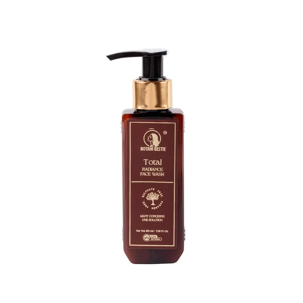
Total Radiance Face Wash — Prevent New PIH at Every Wash
Sulphate-Free · pH Balanced · Salicylic Acid · Niacinamide · Turmeric
Every new breakout is a future dark mark. Salicylic Acid (BHA) — oil-soluble, penetrates the pore lining to dissolve the sebum and debris that block follicles and feed active acne. Fewer active spots mean fewer inflammation events, which means fewer new PIH marks forming. Niacinamide — even at the rinse-off concentration of a face wash, works on the skin during contact to inhibit inflammatory signalling at the follicle opening, reducing the severity of each breakout that does occur. Turmeric (Curcumin) — documented anti-inflammatory and mild tyrosinase-inhibiting activity, supporting both acne control and the first layer of PIH prevention at the surface.
Sulphate-free and pH-balanced at 5.5 — critical because AHAs, Vitamin C, and Retinoids all require an acidic skin environment to function. A high-pH sulphate cleanser raises the skin's pH after washing, reducing the efficacy of every active you apply for the next 30+ minutes. Starting with a correctly pH-balanced wash means your brightening routine can work at full efficacy immediately after cleansing.
Shop Total Radiance Face Wash →The Full PIH Routine — Morning and Evening
Building a consistent routine around proven actives
Morning
- Cleanser — pH-balanced, sulphate-free (Total Radiance Face Wash)
- Vitamin C serum — 10–20% L-Ascorbic Acid or stable derivative; apply to damp skin
- Niacinamide — 5–10% after Vitamin C (wait 60–90 seconds for Vitamin C to absorb)
- Moisturiser — hydration before SPF improves SPF film evenness
- SPF 50+ PA++++ — last step, non-negotiable
Evening
- Cleanser — double-cleanse if wearing SPF or makeup
- AHA (Glycolic or Lactic Acid) — 2–3× per week on alternating evenings
- Retinoid (Adapalene 0.1% or Retinol) — 2–3× per week on evenings alternating with AHA
- Alpha Arbutin or Tranexamic Acid — on non-exfoliant evenings
- Moisturiser — especially important on retinoid nights
Introduce one new active at a time (2 weeks apart) to isolate any sensitivity response. All new actives start at 2–3× per week before daily use.
Still Not Sure What You're Dealing With?
PIH, melasma, solar spots, and acne scars often co-exist on the same face — and require different treatments applied in the right sequence. A dermatologist assessment — including Wood's lamp pigmentation depth testing and scar grading — gives you a specific diagnosis and a prioritised protocol. Most people trying to self-treat hyperpigmentation in India are using the right ingredients for the wrong condition. One consultation corrects the approach and prevents months of ineffective product cycling.
Book Free Consultation → WhatsApp UsFrequently Asked Questions

The Bottom Line — Diagnosis First, Then the Right Treatment
The flat brown mark left by a pimple is PIH. The textured indent is a scar. The symmetrical summer-worsening patch is melasma. These are not three names for the same thing — they are three different biological processes that require three different protocols. Using a scar-filling serum on PIH achieves nothing. Using a brightening serum on an ice pick scar achieves nothing. And using either without daily SPF 50+ PA++++ undoes both.
Most Indian skin care failures in the hyperpigmentation category are not product failures — they are diagnosis failures. The right products, applied to the correctly identified condition, with consistent sun protection, produce measurable, predictable improvements. The timeline is weeks to months, not days — but it is reliable when the biology is correctly addressed.
Start with the touch test. Identify what is flat (pigmentation) and what has texture (scar). Match the treatment to the biology. Protect from UV every single morning without exception. If the diagnosis is unclear or the condition is not responding — a dermatologist assessment with Wood's lamp testing gives you the specific answer in one session and saves months of trial and error.
Shop Total Radiance Face Wash → Free Skin Consultation"A brightening serum for a scar, and a scar filler for PIH — both useless. Diagnosis is the treatment."
Ready to make the honest switch?
Our products are built on the same principles as every article we write — pure ingredients, real results, nothing to hide.
Explore Our Products